
Clinical Journal 2: the anatomy of the skull and spine
My second week brought together several subjects that will form the foundation of my medical education: anatomy, kinesiology, physical education, physiology, an introduction to practical skills, ethics, and pathophysiology.
For someone interested in longevity, prevention, and medical rehabilitation, the connections between these subjects are particularly important.
Understanding the skeletal system, how it contributes to movement, how it interacts with surrounding structures, and what happens when its function is compromised provides a much more comprehensive perspective.
This week, two areas especially caught my attention: the anatomy of the skull and spine, and the terminology we use to describe movement.
I. The Skull
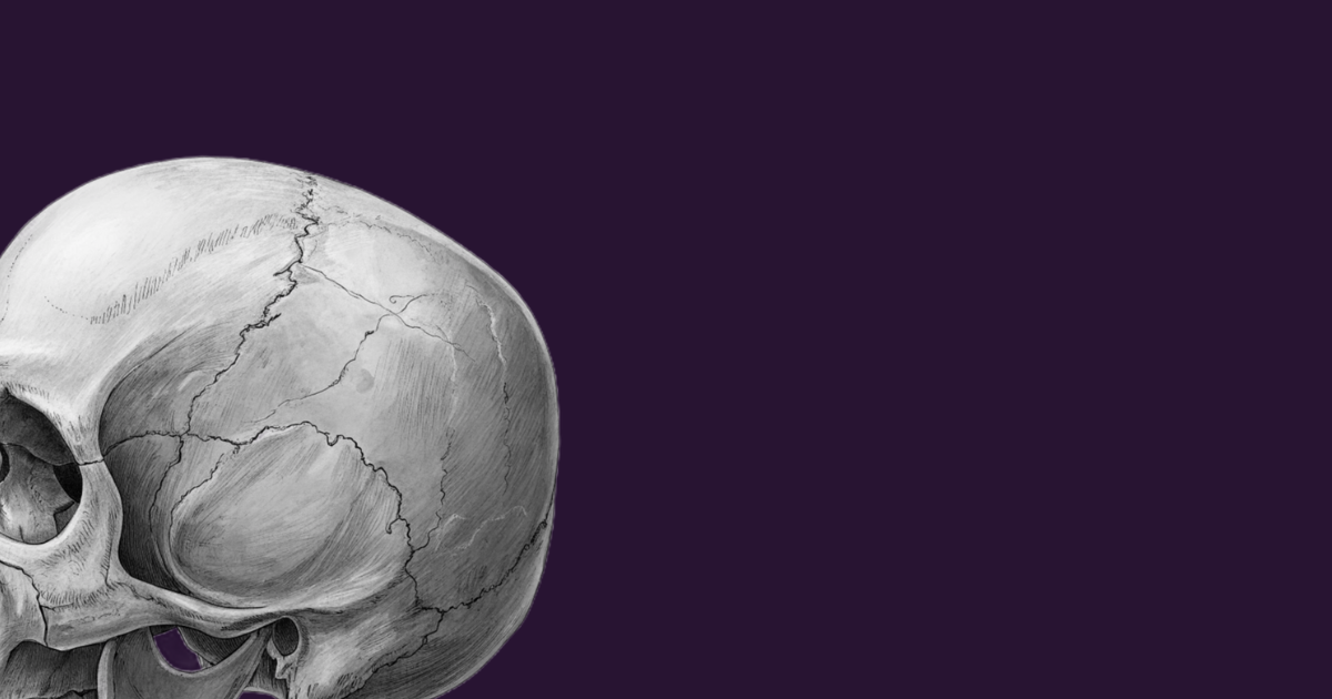
The adult skull is conventionally described as consisting of 22 bones: 8 forming the neurocranium, which encloses and protects the brain, and 14 forming the viscerocranium (facial skeleton). The 6 auditory ossicles and the hyoid bone are generally considered separately from these 22 bones.
The neurocranium encloses and protects the brain, while the viscerocranium forms the bony framework of the face and supports structures involved in functions such as breathing, eating, and vision. The bones of the skull are connected predominantly by sutures, which are fibrous joints that allow little to no movement in the adult.
These connections provide structural continuity while reflecting the developmental processes through which the skull forms. Learning about the skull also means learning how to orient oneself anatomically. Anterior, posterior, superior, inferior, and lateral views reveal different relationships between bones, openings, processes, and other anatomical landmarks.
These landmarks are not simply details to memorize; they help us describe locations precisely and understand the relationships between anatomical structures. The skull is also functionally connected to the rest of the musculoskeletal system.
The mandible articulates with the temporal bones at the temporomandibular joints (TMJs), which allow the movements required for chewing and contribute to the movements involved in speaking. The skull articulates with the first cervical vertebra, the atlas, at the atlanto-occipital joints, which contribute to movements such as nodding the head.
The atlanto-axial joints, between the Atlas (C1) and the Axis (C2), account for a substantial proportion of the rotation involved in turning the head from side to side.
These movements depend on coordinated interactions between joints, muscles, ligaments, and the nervous system. Understanding the relationships between the skull, jaw, and cervical spine provides a foundation for examining head and neck movement. It also establishes a vocabulary for discussing posture, mobility, and musculoskeletal function.
However, anatomical relationships alone cannot explain every symptom. Headaches, jaw pain, and neck pain can have multiple causes. A sound clinical assessment must consider the person’s history, symptoms, examination findings, and relevant differential diagnoses rather than assuming that a structural relationship alone explains the problem.
For now, the key lesson is that anatomical knowledge provides a foundation for understanding movement, assessing function, and approaching clinical problems more systematically.
II. The Spine and the Spinal Cord
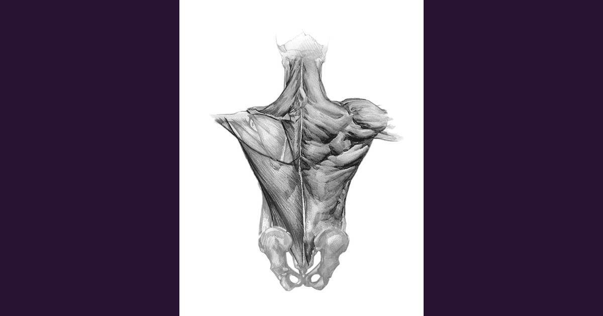
My first encounter with actual vertebrae from the cervical, thoracic, and lumbar regions made another distinction particularly tangible: the difference between the vertebral column and the spinal cord.
The vertebral column is a bony and cartilaginous structure that supports the trunk, helps transmit loads, permits movement, and protects the spinal cord within the vertebral canal.
The spinal cord, by contrast, is nervous tissue that forms part of the central nervous system. It carries signals between the brain and the body and, through its connections with peripheral nerves, contributes to sensation, voluntary movement, reflex activity, and autonomic regulation.
The two structures occupy closely related anatomical spaces, but they perform different functions.
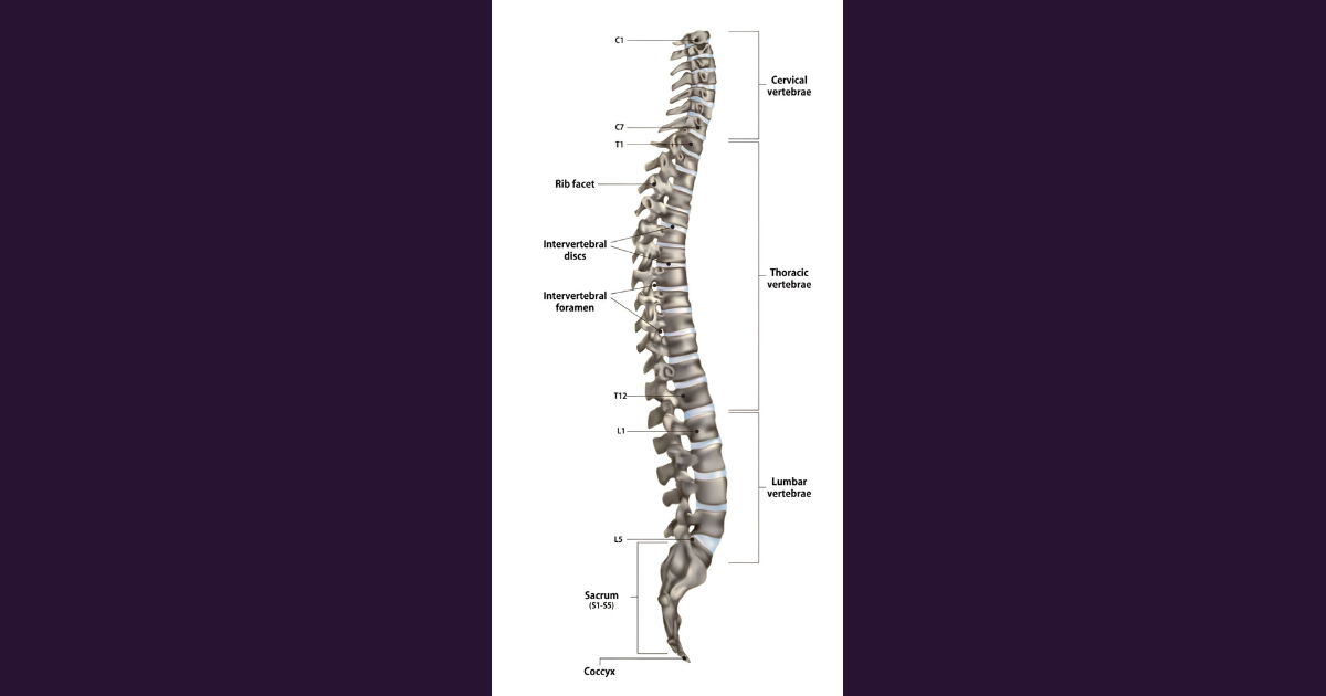
The regions of the vertebral column
The vertebral column is divided into five regions:
| Region | Typical number of vertebrae | General role |
| Cervical | 7 | Supports the head and allows substantial neck mobility |
| Thoracic | 12 | Articulates with the ribs and contributes to the stability and mechanics of the thorax |
| Lumbar | 5 | Bears substantial loads and permits movements of the lower trunk |
| Sacral | 5 | Transmits loads between the vertebral column and pelvis |
| Coccygeal | Usually 4 | Forms the coccyx and provides attachment points for surrounding structures |
The vertebral column is commonly described as developing from 33 – 34 vertebrae.
In adults, the 24 presacral vertebrae remain separate, while the five sacral vertebrae fuse to form the sacrum and the coccygeal vertebrae usually fuse to form the coccyx. The number of coccygeal vertebrae varies between individuals.
The regions differ in shape, size, and function. Cervical vertebrae are adapted to the demands of head and neck movement. Thoracic vertebrae articulate with the ribs, while lumbar vertebrae have large bodies suited to load-bearing.
What does a vertebra do?
Although vertebrae share a general organizational plan, their individual features vary by region.
A typical vertebra includes a vertebral body, a vertebral arch, a vertebral foramen, and several processes (bony projections or extensions that stick out from the main body or arch of the bone).
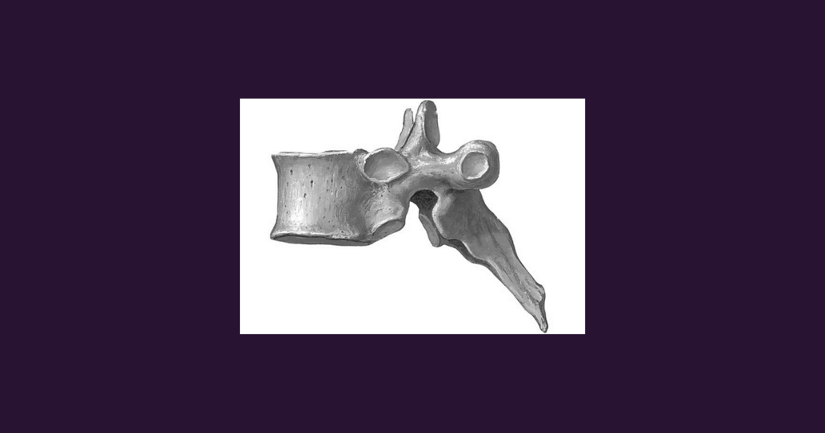
The vertebral body contributes to load-bearing. The vertebral arch surrounds the vertebral foramen, and the successive vertebral foramina form the vertebral canal, which contains the spinal cord and associated structures. The transverse and spinous processes provide attachment points and leverage for muscles and ligaments, while the articular processes form joints with neighbouring vertebrae.
The thoracic vertebrae I encountered during the practical session belong to a region characterized by its articulations with the ribs.
Together, the thoracic vertebrae, ribs, sternum, and associated structures form the thoracic cage, which protects organs such as the heart and lungs while allowing the movements required for breathing.
This is an important example of anatomy as an integrated system: a vertebra contributes not only to the structure of the spine but also to the mechanics of the thorax.
Intervertebral discs
Between most adjacent vertebral bodies are intervertebral discs. These fibrocartilaginous structures help distribute compressive loads and permit movement between vertebrae.
Each disc consists of an outer annulus fibrosus and an inner nucleus pulposus. Together, these components allow the disc to deform under load and contribute to the mechanical behaviour of the vertebral column. The discs work together with the facet joints, ligaments, muscles, and vertebral bodies. No single structure is responsible for the spine’s overall stability or mobility.
This matters because back pain is often discussed as if it must originate from one identifiable structure. In reality, spinal pain can involve multiple mechanisms, and imaging findings do not always correspond directly to symptoms. Structural knowledge is essential, but it must be combined with clinical assessment.
The spinal cord and spinal nerves
The spinal cord runs within the vertebral canal, but in most adults it terminates around the L1–L2 vertebral level, with individual variation.
Below this point, the lumbar and sacral nerve roots descend through the canal as part of the cauda equina before exiting through the appropriate intervertebral foramina.
There are typically 31 pairs of spinal nerves:
- 8 cervical pairs
- 12 thoracic pairs
- 5 lumbar pairs
- 5 sacral pairs
- 1 coccygeal pair
These nerves carry sensory information toward the central nervous system and motor signals toward muscles. They also contain autonomic fibres where applicable.
Damage to the spinal cord, a spinal nerve root, or a peripheral nerve can produce different patterns of neurological impairment.
Understanding their locations and functions provides the foundation for learning how clinicians assess weakness, sensory changes, reflexes, and other neurological findings.
Why the spine matters in rehabilitation and longevity
The vertebral column is central to posture, movement, load transfer, and the protection of neural structures. Its function depends on the interaction of bones, discs, joints, ligaments, muscles, and the nervous system.
In rehabilitation, the objective is not simply to make a structure look anatomically correct.
It is to understand how a person moves, what limits their function, and which interventions may help them perform meaningful activities more safely and independently.
This connects directly to a broader understanding of longevity. Living longer is one dimension of health; maintaining the ability to move, work, participate, and remain independent is another.
The World Health Organization defines rehabilitation as a set of interventions designed to optimize functioning and reduce disability in people with health conditions, taking into account their interaction with the environment.
Rehabilitation is relevant not only after an injury but also in the context of disease, chronic conditions, and age-related functional decline.
III. Anatomical Movements
Anatomy helps us identify the structures involved in movement, while kinesiology helps us understand how those structures work together to produce it.
The scientific study of human movement draws on several related disciplines, including kinesiology and biomechanics.
Kinesiology examines human movement from a broad perspective, while biomechanics focuses on the mechanical principles underlying movement, including forces, motion, and the interactions between the body and its environment.
Biomechanics is often considered a subfield of kinesiology, although the relationship between the two disciplines can vary across academic contexts.
The term kinesiology comes from the Greek words kinesis (movement) and logos (study).
Movement terminology can initially feel like a long list of definitions. Yet each term provides a precise way to describe what happens at a joint or between body segments. The reference point is the anatomical position: standing upright, facing forward, with the arms at the sides, the palms facing forward, and the thumbs pointing laterally.
Anatomical movements are described using this standardized reference position, together with the relevant planes and axes of movement.
The Three Anatomical Planes
The body is commonly described in relation to three anatomical planes:
- Sagittal plane: divides the body into right and left portions. Flexion and extension commonly occur in this plane.
- Frontal (coronal) plane: divides the body into anterior and posterior portions. Abduction and adduction commonly occur in this plane.
- Transverse (horizontal) plane: divides the body into superior and inferior portions. Rotational movements commonly occur in this plane.
These planes provide useful reference systems rather than rigid descriptions of every movement.
Many everyday activities and sporting actions involve movement across multiple planes and the coordinated action of several joints.
Fundamental movements
Understanding anatomical movement terminology begins with a few fundamental movements that describe how body segments move in relation to one another.
Flexion and extension describe changes in the angle between body segments at a joint. Flexion generally decreases the angle, as when bending the elbow, while extension increases the angle or straightens the joint, as when straightening the elbow.
Abduction and adduction describe movement away from and toward the body’s midline, respectively.
Raising the arm sideways is an example of abduction, while bringing it back toward the body illustrates adduction.
Internal and external rotation describe the rotation of a body segment around its longitudinal axis, toward or away from the midline, according to the joint and anatomical reference. For example, rotating the thigh inward or outward at the hip demonstrates internal and external rotation.
Circumduction is a circular movement that combines flexion, abduction, extension, and adduction in sequence. Making a circle with the arm is a familiar example.
Lateral flexion refers to bending the neck or trunk to one side, as when tilting the head toward one shoulder or bending the trunk sideways.
Other terms describe more specialized movements.
Pronation and supination describe the rotation of the forearm that turns the palm downward or upward, respectively, in the anatomical reference position.
Dorsiflexion and plantar flexion describe movements at the ankle. Dorsiflexion brings the top of the foot toward the shin, while plantar flexion points the foot downward, away from the shin.
Inversion and eversion describe movements of the foot in which the sole turns inward or outward, respectively.
Elevation and depression describe upward and downward movement of a body part. Raising and lowering the shoulders are common examples.
Protraction and retraction describe forward and backward movement of a body part.
These terms are commonly used when discussing the scapula and mandible, such as when the shoulders move forward or the jaw moves forward and backward.
The fingers also have specific movement terminology.
Abduction and adduction describe spreading the fingers apart and bringing them together, respectively, using the third digit, or middle finger, as the reference line.
The thumb has a different anatomical orientation and therefore requires special consideration when describing its movements. Learning these terms creates a common language for describing movement. Rather than simply saying that a person “cannot move the arm properly,” a clinician can begin to specify which movement is restricted, at which joint, under what conditions, and to what extent.
Muscle Contractions: Movement is Not Always Visible
Another important distinction concerns how muscles generate force.
Three commonly described types of muscle action are concentric, eccentric, and isometric.
Concentric action: the active muscle shortens while producing force. An example is the upward phase of a biceps curl, when the elbow flexes against a load.
Eccentric action: the active muscle lengthens while producing force. During the controlled lowering phase of the same curl, the elbow flexors work eccentrically to control the descent.
Isometric action: the muscle produces force without appreciable change in the overall length of the muscle–tendon unit. Holding a position against a load is a familiar example.
More about Anatomical Movements: https://www.youtube.com/watch?v=r0pVf60fTIA
IV. What Medical Education Could Learn from Business
Moving between business and medicine naturally invites comparisons between the two educational environments.
One idea I have carried with me from my business education and experience across various sectors is the importance of focusing on growth, rather than on failure alone.
In business, managing risk is essential, but it is not the same as building the capacity for growth.
Identifying and reducing risks matters, yet so does creating the conditions for development and long-term progress.
I wonder whether a similar distinction is useful when thinking about health.
Preventing disease and managing complications are essential.
But could we also give greater attention to maintaining function, building physical capacity, supporting healthy ageing, and helping people preserve their independence before significant limitations emerge? This is not an argument against disease prevention or treatment. Nor is it a claim that medicine neglects healthy functioning; many medical and public health disciplines already address it.
Rather, it is a question of emphasis: how can health systems balance the prevention and management of disease with a more explicit focus on long-term function and well-being? For someone interested in rehabilitation and longevity, this question feels particularly relevant.
The business perspective also encourages systems thinking. Outcomes rarely depend on a single variable.
In an organization, performance emerges from the interaction of people, incentives, processes, resources, and decisions.
Similarly, human function emerges from the interaction of biological systems and is shaped by behaviour and the surrounding environment.
The analogy has its limits, of course. A human being is not a company, and biological systems cannot be managed as if they were simple organizational processes.
Nevertheless, systems thinking can offer a useful framework for exploring how different factors interact and influence outcomes.
I am still at the beginning of my medical education, so I would not yet draw broad conclusions about how medical curricula address prevention and the maintenance of function. For now, this is a question I hope to explore further as my understanding of medicine develops.
V. What Business Education Could Learn from Medicine
The comparison works in the other direction, too.
One aspect of medical education I find particularly valuable is the opportunity to connect theoretical knowledge with anatomical structures, practical skills, and, eventually, clinical practice.
Even a first encounter with an anatomical specimen can change how a concept is understood.
I believe business education can benefit from a similar principle: earlier and more sustained exposure to the environments in which knowledge must actually be applied.
Students can learn frameworks, models, and theories, but the meaning of these concepts often becomes clearer when they encounter real people, practical constraints, competing priorities, and consequential decisions.
Direct exposure can reveal the gap between a well-constructed model and the complexity of a real-world situation.
This is not to suggest that every medical course provides sufficient practical experience from the outset, or that business education universally lacks it.
Both fields combine theory, simulation, practice, and supervised experience in different ways. The important question is how effectively these elements are integrated.
My own experience so far has also prompted me to reflect on teaching methods.
Some sessions have relied heavily on text-dense slides and dictation. This is an early observation, not a definitive assessment of the curriculum or its effectiveness.
Lectures and note-taking can play a useful role in learning, particularly when students need to establish a foundation of terminology and factual knowledge.
However, I wonder how effectively that foundation is reinforced through active recall, practical demonstrations, discussion, feedback, and opportunities to apply concepts to real situations. In a subject such as anatomy, memorizing the names of vertebral structures is one task.
Identifying those structures on a specimen, understanding their anatomical relationships, and explaining their functional significance represent different levels of learning.
The same distinction applies in business: knowing a model is not the same as recognizing when it applies, when its assumptions no longer hold, and how it might need to be adapted to a particular context.
I would like to see these dimensions of education considered together: theoretical understanding, practical exposure, and the ability to think critically and independently.

VI. From Business Suit to Medical Scrubs. And Back Again.
Business buildings, hospitals, university classrooms, and back to business.
For me, moving between these environments is not merely a logistical transition; it is a psychological one, too. Each shift requires me to adapt to a different role while keeping sight of the broader purpose behind the journey.
Each environment has its own vocabulary, expectations, standards, and ways of evaluating competence. In business, I learned to think in terms of systems, strategy, and growth. Medical education introduces a different language: anatomy, physiological mechanisms, clinical reasoning, and the responsibility that comes with working in a field concerned with human health.
Some of these perspectives complement one another. Others challenge me to reconsider assumptions that once felt familiar.
The challenge is to remain open to learning without rushing to conclusions. I am learning to become comfortable with not knowing, to build my knowledge carefully, and to recognize when a question requires more evidence than I currently have.
That may be one of the most important psychological adjustments in this journey: accepting that competence develops incrementally, and that intellectual curiosity must be accompanied by discipline and humility.
The phrase “from the business suit to medical scrubs and back again” captures the contrast between these professional worlds.
More importantly, it reminds me that I am learning to move between different ways of understanding problems, while recognizing that medicine carries responsibilities that cannot be reduced to efficiency, performance, or business outcomes.
VII. What I Am Taking Away from the 2nd WeeK
This week went beyond anatomical terminology.
It gave me my first hands-on encounter with vertebrae and introduced me to the language used to describe movement. It also began to show me how anatomy, physiology, and kinesiology provide complementary foundations for understanding human function.
The connections to rehabilitation are already apparent: anatomical structures support movement, the nervous system coordinates it, and clinical assessment helps us understand where function is limited and what might be improved. The connections to longevity are broader and will require further study.
Still, the idea that health involves not only preventing disease but also maintaining the ability to function, participate in life, and preserve independence is a perspective I want to explore throughout this journey.
At the same time, I am beginning to reflect on education itself: how knowledge is taught, how theory becomes practice, and what medicine and business might learn from each other without overlooking their distinct purposes and responsibilities.
For now, my task is to build a solid foundation: to learn the structures, understand the mechanisms, and connect them to function.
The larger questions surrounding prevention, rehabilitation, and longevity will become more meaningful as that foundation develops.
Until we meet again,
T.
CONTINUE YOUR JOURNEY

The third week was largely about learning to see what cannot be observed directly: cells and their components, the electrical activity of the heart, the concentration of a [...]
-
My second week brought together several subjects that will form the foundation of my medical education: anatomy, kinesiology, physical education, physiology, an introduction to practical skills, ethics, and [...]

Today I launch the first Clinical Journal post. This is the beginning of a three-year journey into formal medical education and a way to document my clinical [...]

Lorem ipsum Lorem ipsum dolor sit amet, consectetur adipiscing elit, sed do eiusmod tempor incididunt ut labore et dolore magna aliqua. Ut enim ad minim veniam, quis nostrud [...]

Lorem ipsum Lorem ipsum dolor sit amet, consectetur adipiscing elit, sed do eiusmod tempor incididunt ut labore et dolore magna aliqua. Ut enim ad minim veniam, quis nostrud [...]

Lorem ipsum Lorem ipsum dolor sit amet, consectetur adipiscing elit, sed do eiusmod tempor incididunt ut labore et dolore magna aliqua. Ut enim ad minim veniam, quis nostrud [...]
Lorem ipsum Lorem ipsum dolor sit amet, consectetur adipiscing elit, sed do eiusmod tempor incididunt ut labore et dolore magna aliqua. Ut enim ad minim veniam, quis nostrud [...]