
Clinical Journal 3: Making the Invisible Visible
The third week was largely about learning to see what cannot be observed directly: cells and their components, the electrical activity of the heart, the concentration of a solution, and the anatomy of the upper limb.
The connecting idea was measurement.
Whether through a microscope, an electrocardiograph, laboratory equipment, or anatomical observation, medicine depends on methods that transform hidden structures and processes into interpretable information.
I. Seeing cells: from light to electrons
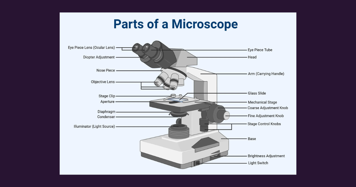
In cell biology, I studied the microscope: its components, its use, and the different ways it can reveal cellular structure.
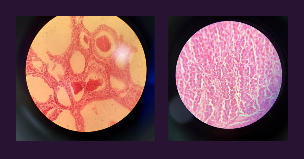
The light microscope uses visible light to form an image. Because native animal cells are largely colourless and transparent, specimens are commonly fixed and stained to increase contrast.
A stain appears coloured because it selectively absorbs certain wavelengths of visible light, altering the spectrum of light transmitted through or reflected by the specimen.
The resolving power of a light microscope is limited by the wavelength of light and by the numerical aperture of the objective lens. Under ideal conditions, a conventional light microscope can distinguish two points separated by approximately 0.2 µm.
Phase-contrast microscopy allows living, unstained cells to be examined by converting differences in optical path length into differences in brightness.
Other optical approaches, such as dark-field and differential interference contrast microscopy, use different ways of generating contrast.
Fluorescence microscopy adds another layer of specificity.
Fluorophores absorb light at one wavelength and emit light at a longer wavelength. With appropriate filters, researchers can label and visualize particular molecules or cellular structures.
Fluorescence greatly improves contrast and specificity, but conventional fluorescence microscopy remains constrained by diffraction; super-resolution techniques are required to push resolution toward the nanometre scale.
Confocal microscopy uses laser scanning and optical sectioning to build sharper fluorescent images, including three-dimensional reconstructions.
Polarized-light microscopy can highlight ordered, fibrillar, or linear structures such as collagen, myofibrils, and myelin.
The electron microscope works differently. Instead of visible light, it uses a beam of accelerated electrons focused by electromagnetic lenses. Because electrons have much shorter effective wavelengths than visible light, electron microscopy can reveal ultrastructure macromolecular complexes and organelles far below the limit of conventional light microscopy.
Transmission electron microscopy produces images from electrons passing through ultrathin specimens, while scanning electron microscopy produces surface-focused images.
Cryo-electron microscopy allows vitrified, hydrated specimens to be examined without conventional chemical fixation or staining, helping preserve structures close to their native state.
What stayed with me was the realization that every method reveals a particular version of reality.
A stained optical image, a fluorescent label, and an electron-microscopy image do not show the same thing in the same way.
Choosing a method means choosing what question can be asked.
II. The cardiovascular system: structure, pump, and signal
In physiology, we began studying the cardiovascular system and the use of the electrocardiograph.
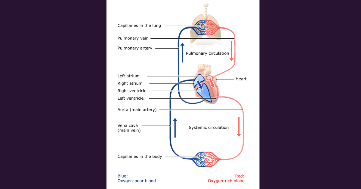
The heart is a muscular, four-chambered pump. It receives blood, generates pressure, and propels it through two circuits: the pulmonary circulation, which carries blood between the heart and lungs, and the systemic circulation, which delivers blood to the rest of the body.
The wall of the heart has three main layers: the epicardium, myocardium, and endocardium. The pericardium is the surrounding sac that encloses the heart.
The myocardium is the contractile muscle layer. The heart also contains specialized tissue responsible for initiating and conducting electrical impulses.
The sinoatrial node, atrioventricular node, atrioventricular bundle, bundle branches, and Purkinje fibres form the cardiac conduction system. The heart’s activity can be described through electrical, mechanical, and acoustic manifestations.
Electrical activation precedes mechanical contraction; the electrocardiogram records the electrical part of this sequence.
The electrocardiogram
An electrocardiogram, or ECG, is a graphical recording of the heart’s electrical activity detected at the body surface. It does not measure contraction directly. Rather, it records the summed electrical potentials generated as cardiac cells depolarize and repolarize.
During depolarization, the electrical potential across the cardiac cell membrane becomes less negative and may briefly become positive on the inside relative to the outside.
Repolarization restores the resting distribution of charge.
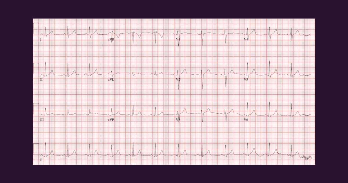
These electrical events spread through the heart in an organized sequence, beginning normally in the sinoatrial node and continuing through the atria, atrioventricular node, His bundle, bundle branches, Purkinje fibres, and ventricular muscle.
The standard clinical ECG uses 12 leads: three bipolar limb leads, three augmented limb leads, and six precordial chest leads. The limb leads provide a frontal-plane view of cardiac electrical activity, while the precordial leads provide a transverse-plane view.
A lead is not simply an electrode. It represents the difference in electrical potential between two points, with a defined positive and negative orientation.
Because the heart’s electrical axis has a three-dimensional orientation, different leads capture different projections of the same underlying activity.
Interpreting an ECG involves assessing rate and rhythm, the cardiac electrical axis, the P wave, QRS complex, T wave.
Each component reflects a different aspect of cardiac electrical activation or recovery.
Electrical changes occurring at the level of individual cardiac cells become a pattern on paper or a screen.
That pattern can then be used to evaluate rhythm, conduction, and, in clinical contexts, possible pathology.
The cardiovascular system under effort
We also discussed how the cardiovascular system responds to exercise.
During acute exercise, heart rate, stroke volume, and cardiac output increase to meet the metabolic demands of active muscle.
Cardiac output (the product of heart rate and stroke volume) can increase approximately five- to six-fold during maximal effort.
In highly trained endurance athletes, maximal cardiac output may exceed 30 L/min.
Blood flow is also redistributed. Active skeletal muscle receives a greater proportion of cardiac output, while vascular resistance in other beds may change to support this redistribution.
Systolic blood pressure generally rises during dynamic exercise, while diastolic pressure may remain similar or even decrease because of vasodilation in contracting muscle.
With endurance training, the cardiovascular system adapts. Resting heart rate often decreases, stroke volume increases, and sustained training may lead to physiological cardiac remodelling, including changes in ventricular chamber size and myocardial mass.
These adaptations can support a larger stroke volume and greater maximal cardiac output. This is directly relevant to rehabilitation and prevention.
Exercise is not only a way to expend energy; it is a physiological stimulus that can reshape cardiovascular function over time. The clinical question is always one of dose, context, individual capacity, and safety.
III. Measurement in the laboratory
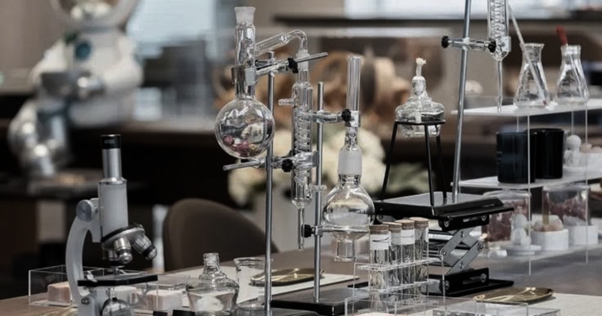
In biochemistry, we discussed laboratory glassware, measuring instruments, and the calculation of percentage concentrations.
The equipment itself may seem basic: beakers, Erlenmeyer flasks, graduated cylinders, volumetric flasks, pipettes, burettes, test tubes, balances, pH meters, and spectrophotometers. But each has a specific role.
A beaker is useful for preparing or mixing solutions, but it is not designed for precise volume measurement. A volumetric flask is.
A pipette or burette allows more accurate delivery of liquid volumes. An analytical balance can measure mass to very small increments, while a pH meter and spectrophotometer convert physical properties (hydrogen-ion activity or light absorption) into quantitative information.
The lesson is similar to the one from microscopy: precision depends on using the right instrument for the right question.
In the medical field, a measurement is never only a number. It has an instrument, a method, a context, and a potential source of error.
IV. The upper limb
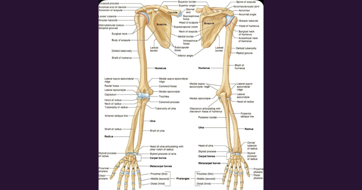
In anatomy class, I studied the skeleton of the upper limb and its joints.
The upper limb consists of the shoulder girdle and the free upper limb. The shoulder girdle (pectoral girdle) comprises the scapula and clavicle and connects the upper limb to the axial skeleton through the sternoclavicular joint.
The free upper limb includes the humerus, radius, ulna, carpal bones, metacarpal bones, and phalanges.
The scapula is a flat, triangular bone on the posterior thorax. Its spine continues laterally as the acromion, which articulates with the clavicle.
The glenoid cavity articulates with the head of the humerus, forming the shoulder joint. The coracoid process provides attachment for muscles and ligaments involved in shoulder stability and movement.
The clavicle is an S-shaped bone between the sternum and scapula. It acts as a strut that keeps the upper limb away from the trunk while allowing considerable mobility.
The humerus articulates with the scapula proximally and with the radius and ulna distally. At the elbow, the trochlea of the humerus articulates with the ulna, while the capitulum articulates with the radius.
The radius and ulna also articulate with each other, enabling pronation and supination of the forearm.
The wrist contains eight carpal bones arranged in two rows. The thumb is particularly notable: the saddle-shaped articulation between the trapezium and the first metacarpal supports opposition, one of the movements that makes precise grasping possible.
The hand contains five metacarpal bones and fourteen phalanges: two for the thumb and three for each remaining finger.
The upper limb is a system built for a remarkable range of functions: reaching, gripping, manipulating, writing, lifting, and communicating.
V. What I take from this week
This week showed me that medicine often begins with a problem of visibility.
Many cellular structures cannot be seen with the naked eye. The electrical activity of the heart cannot be observed directly. The concentration of a solution cannot be reliably determined without measurement.
And the anatomy of the upper limb becomes meaningful when its structures are understood in relation to movement and function.
Microscopy, ECG, laboratory instruments, and anatomical study are different tools, but they share a common purpose: to make complex biological processes observable, measurable, and interpretable.
For rehabilitation and prevention, this matters because function depends on structures and signals that are not always directly visible.
Movement emerges from the interaction of bones, joints, muscles, nerves, metabolism, and the environment.
Understanding it requires both observation and interpretation.
Beyond the scientific content, this week also made me reflect on mathematics.
At school, mathematics is often taught as an isolated subject, with much of the focus placed on memorising formulas and learning abstract concepts. Yet its real value becomes much clearer when we begin to apply it.
Since completing four years of intensive mathematics in high school, I have found it useful in marketing, strategy, and business development. Now, I am beginning to recognise its relevance in medicine as well.
I believe mathematics should be taught from an early stage of education not only as a discipline in its own right, but also through its connections to the world around us, regardless of what students eventually choose to pursue.
Until education makes these connections more consistently, we can still take the initiative to discover them for ourselves.
The world is interconnected, and knowledge rarely exists in isolation. When concepts are learned without being connected to other ideas or real-world applications, they risk becoming disconnected pieces of information, easily forgotten and rarely put to use.
Perhaps this is one of the most valuable lessons I am taking from this week: learning becomes more meaningful when we understand not only individual concepts, but also how they connect, complement, and give meaning to one another.
About Clinical Journal
Clinical Journal documents the development of clinical knowledge, skills, and perspective through formal education and experience in healthcare.
It connects learning about the human body, clinical practice, and patient care with La Syntropie’s broader exploration of prevention and longevity.
The project examines how evidence translates into practice, with particular attention to maintaining health, function, and independence across the lifespan.
Clinical Journal is part of La Syntropie, an interdisciplinary platform exploring longevity at the intersection of science, healthcare, technology, enterprise, and policy.
Until we meet again,
T.
CONTINUE YOUR JOURNEY
-
The third week was largely about learning to see what cannot be observed directly: cells and their components, the electrical activity of the heart, the concentration of a [...]
My second week brought together several subjects that will form the foundation of my medical education: anatomy, kinesiology, physical education, physiology, an introduction to practical skills, ethics, and [...]

Today I launch the first Clinical Journal post. This is the beginning of a three-year journey into formal medical education and a way to document my clinical [...]
Lorem ipsum Lorem ipsum dolor sit amet, consectetur adipiscing elit, sed do eiusmod tempor incididunt ut labore et dolore magna aliqua. Ut enim ad minim veniam, quis nostrud [...]

Lorem ipsum Lorem ipsum dolor sit amet, consectetur adipiscing elit, sed do eiusmod tempor incididunt ut labore et dolore magna aliqua. Ut enim ad minim veniam, quis nostrud [...]

Lorem ipsum Lorem ipsum dolor sit amet, consectetur adipiscing elit, sed do eiusmod tempor incididunt ut labore et dolore magna aliqua. Ut enim ad minim veniam, quis nostrud [...]

Lorem ipsum Lorem ipsum dolor sit amet, consectetur adipiscing elit, sed do eiusmod tempor incididunt ut labore et dolore magna aliqua. Ut enim ad minim veniam, quis nostrud [...]